In a surprising and rare medical case in China, doctors discovered a growing ‘twin sister’ fetus inside the skull of a one-year-old girl.
This strange find has caused delays in her speech development and has drawn significant attention from the medical community and the public.

Unusual symptoms when she was born.
The young girl’s parents first noticed something was wrong when she began to miss developmental milestones, particularly in her speech.
Concerned about her delayed development, they took her to the hospital for a thorough examination. Initially, the doctors conducted several routine tests but found nothing unusual.
According to the little girl’s parents, abnormalities had already been detected before the child’s birth during a 33-week examination.

However, the examination could not identify the exact issue, as the study noted that an MRI “could not provide more information due to space occupation”.
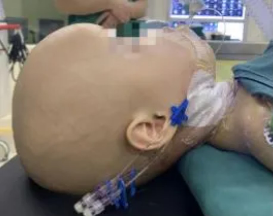
When the baby was delivered four weeks later, she was born with “a large head circumference”. Later on, more concerns arose when she was “only able to say ‘mom'” around the age of one-year-old.
However, they decided to perform a more detailed imaging scan to rule out any hidden issues.
A ‘twin sister’ fetus appeared inside the little girl’s skull.
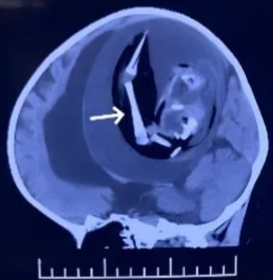
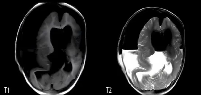
To their astonishment, the scans revealed a mass inside the girl’s skull.
After closer inspection, the medical team identified the mass of a fetus, which had been growing inside her head.
Fetus in fetus
This extremely rare condition, known as “fetus in fetus,” occurs when a malformed fetus is found within the body of its twin.
In this case, the fetus had developed in the girl’s brain, a scenario that is almost unheard of in medical literature.
The doctors immediately understood the urgency of the situation.
The presence of the fetus in the girl’s skull was putting pressure on her brain, which explained her delayed speech development and posed serious risks to her overall health.
The process of removing the fetus is fraught with difficulties
The medical team quickly devised a plan to remove the fetus through a delicate surgical procedure.
The surgery was complex and required a highly skilled team of neurosurgeons.
They carefully navigated the intricate structures of the girl’s brain to reach and remove the fetus without causing additional damage.
The removal of the fetus is expected to alleviate the pressure on her brain, allowing her to develop her speech and other skills normally.
This rare case has sparked widespread interest and discussion among medical professionals.
“Fetus in fetu” is a highly unusual condition, with only about 200 cases reported worldwide.
The fact that the fetus developed in the brain makes this case even more extraordinary.
Researchers and doctors are keen to study this case further to understand how such an anomaly can occur and to learn from the surgical approach taken to treat it.
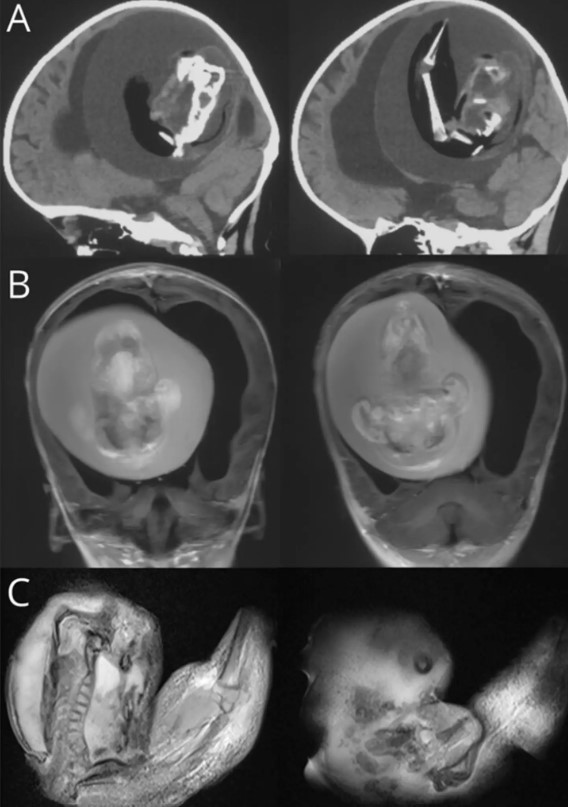
This is truly an extremely rare and heartbreaking case. After the surgery to remove the tumor, the doctors clearly observed the shapes of a fetus, such as the mouth, eyes, arms, and hands.
The little girl passed away after removing the fetus growing inside her skull.
Despite the doctors having conducted thorough examinations, tests, and careful surgical planning, the one-year-old still experienced uncontrollable seizures after the surgery.
Tragically, the child passed away only 12 days later.
The study concluded that while doctors can identify teratomas through anatomical and imaging techniques, surgical removal is the only curative option. However, the prognosis is often poor.